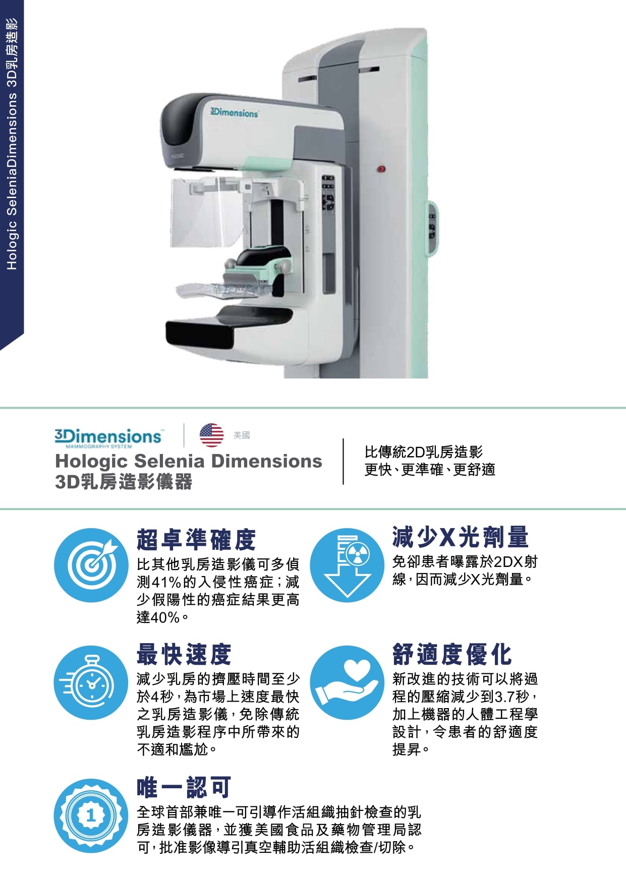

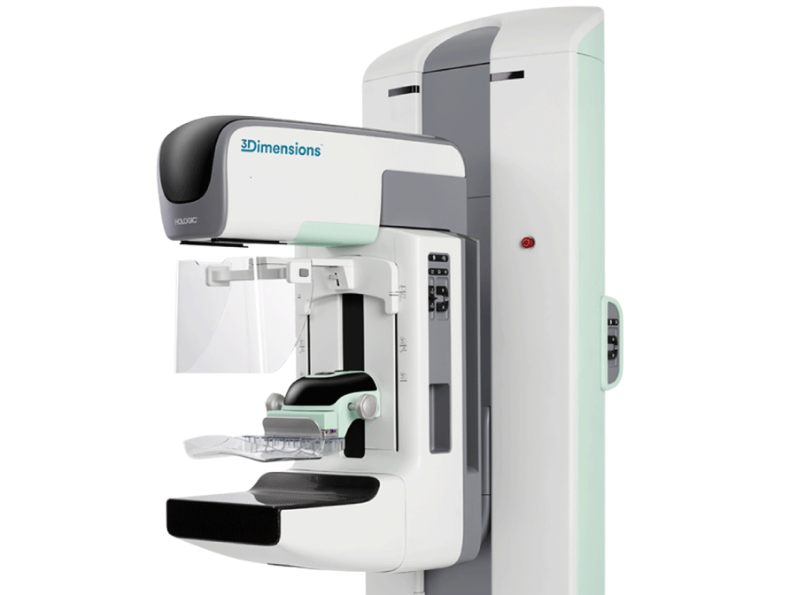
The new 3D Hologic Selenia Dimensions technology reduces the time of breast compression by up to 3.7 seconds. Each breast will be scanned at two different angles, resulting in a total of four sets of images. The entire mammogram takes approximately 20 minutes to complete.
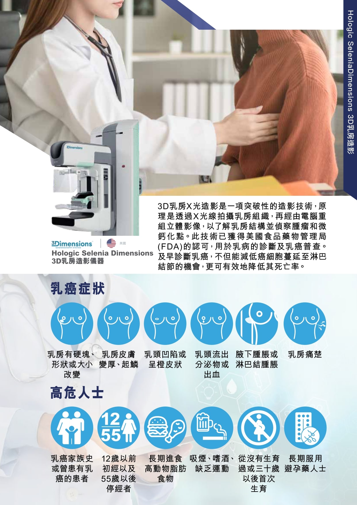
It is recommended that women over the age of 40 undergo routine mammography screening every two years. If you notice any symptoms of breast cancer or have a strong family history of breast cancer, seek help from your healthcare provider and schedule an appointment for a screening mammogram. Early detection of breast cancer can cure and save lives.
It will be more painful for pregnant women to conduct mammography while pregnant as they have more swollen breasts and dense mammary glands than normal women. Therefore, an ultrasound scan is currently the preferred method of breast screening during pregnancy.
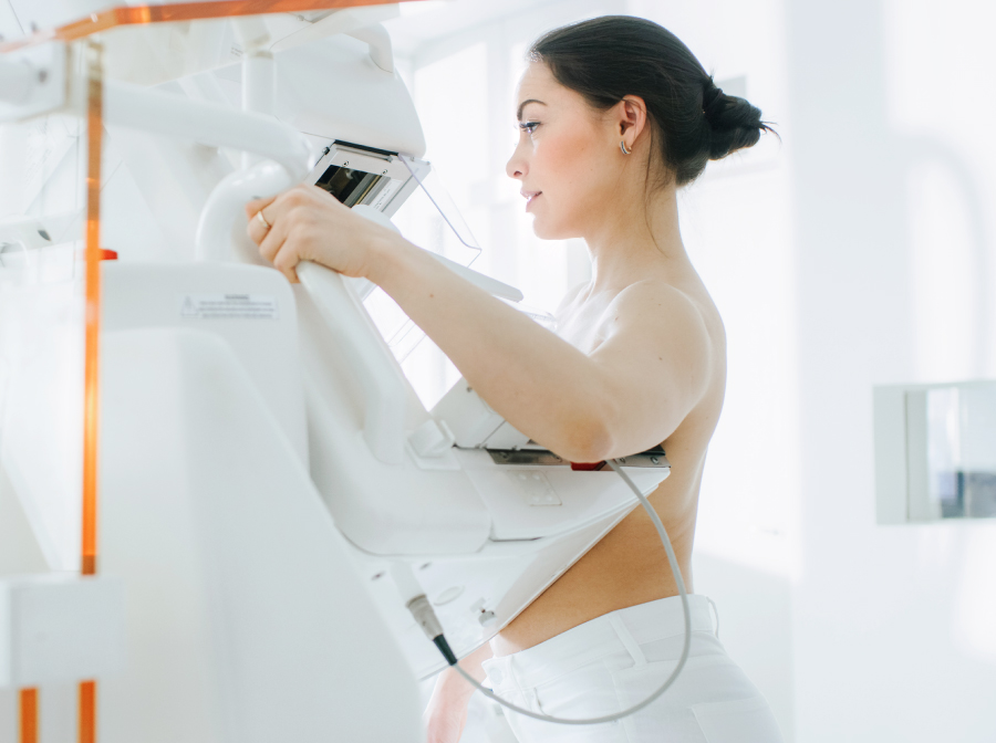
Mammography utilises X-ray contrast and high-resolution films to visualise the internal conditions of the breasts. The breasts need to be pressed to secure them in place while dispersing the internal tissue for mammography. The reason why having the traditional 2D or early 3D mammography in the past is painful is because of the lack of accessories and the prolonged pressing time. The new and improved technology is equipped with accessories to reduce the compression time to 3.7 seconds. The ergonomic considerations of the machines are designed to improve patients' comfort, some patients even give comments on their painless 3D Mammography experience as well.
Before the mammography, you will need to take off your clothes for the upper part of the body and the bra and change into the clothes that we provided. You will need to take off your clothes for the upper part of the body during the mammography. A female radiologist will accompany you throughout the entire procedure. She will place your breast on a plate for the examination. Each breast will be scanned at two different angles, resulting in a total of four sets of images.