



【CT, MRI, Xray-There are many options for image inspection, introduction to FAQ】
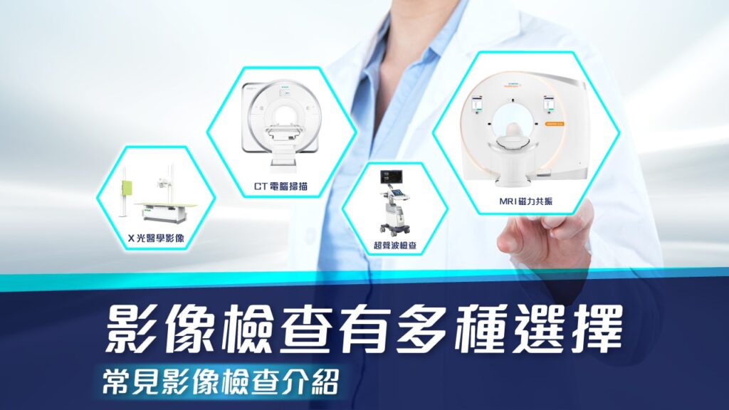
Imaging examination is a medical examination method that uses different types of radiation or energy to obtain image information of the human body's internal tissue structure, function, blood flow, and lesions. These images can provide the information doctors need to diagnose, monitor conditions and plan treatment.
Technology continues to advance, and there are more and more options for imaging examinations. Different examination tools are good at diagnosing different diseases and have different applications. The following is a brief introduction to common imaging inspection tools. The advantages and disadvantages of each inspection are clear at a glance. (This is just a basic introduction and will not go into detail about the physical principles behind each examination. For practical application decisions, please follow the opinions of experts and radiographers)
X-ray
●Advantages
Convenient, fast and low cost
Highly valuable in assessing the integrity and overall structure of bones
●Disadvantages
The imaging quality of soft tissue is not good, so it is basically impossible to see structures such as muscles/tendons/ligaments.
There is radiation, but the amount is very low
When to use
Because X-ray can obtain results quickly and has good diagnostic value for fractures, it is often used as the first line of sports injuries and trauma. Generally speaking, if you want to do imaging examination, you will almost always have an X-ray first.
CT (computer scan)
●Advantages
The bone structure can be obtained completely, and it can be analyzed from different sections and even reconstructed in 3D. In the presence of contrast agent, it can also provide information on some soft tissues (tumors, etc.)
The inspection is fairly fast and usually takes about a few minutes to complete.
●Disadvantages
The cost is relatively high, and the amount of radiation is relatively large compared to X-ray.
Without the use of developer, the imaging of soft tissue is still not detailed enough.
When to use
Usually after the fracture is determined to be diagnosed and treated, CT may be used as a pre-diagnosis and treatment planning, because CT can provide more information about the bone structure. In addition, in certain diseases such as femoroacetabular impingement, CT also has great value in preoperative diagnosis and postoperative evaluation.
Ultrasound
●Advantages
The inspection is convenient (it is portable) and the price is relatively cheap
Dynamic scanning allows you to see real-time images
Good imaging quality of soft tissue
Has both diagnostic and palliative value
No radiation
●Disadvantages
Inspection results will vary greatly depending on the examiner's skill
Poor bone penetration
Easily shielded by air and tissue and unable to see underlying structures
When to use
Ultrasound is now one of the most widely used examination tools. The main reason is that it can find many soft tissue injuries, and many treatments can be completed under the guidance of ultrasound. Although ultrasound cannot penetrate bones, it actually plays a role in diagnosing very early or subtle fractures. Ultrasound can be said to be the first test done after X-ray.
MRI
●Advantages
Can provide complete soft tissue, bone and joint information
Soft tissue imaging results are the best among all examinations
No radiation
●Disadvantages
Relatively few configurations, usually only available in large diagnostic centers or hospitals
The price is high, the examination takes a long time, and it is difficult for patients to cooperate
Keep it still for about half an hour or even longer, otherwise the image quality may be affected.
When to use
MRI is often used to obtain more soft tissue information. For example, today ACL rupture (anterior cruciate ligament rupture) can be determined through physical examination. The main reason for arranging MRI is to see if there are other structural damages, because this may affect the treatment plan. MRI will have better diagnostic value for stress fractures, early bone edema, labral tears, osteochondral lesions, etc.
Clinical medical imaging not only expands the scope of human body examination and improves the level of diagnosis, but also enables treatment of certain diseases and has become an important pillar in medical work.