




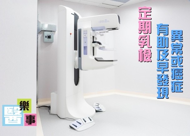
Last month, Miss B came to my diagnostic center for a check-up. Ms. B is unmarried and 45 years old. She found a hard lump under her left armpit while taking a shower a week ago, so she went to see a doctor. The doctor immediately referred her for mammogram and ultrasound after examination. I chatted with her for a while and found out that she had never had any breast-related examinations, there was no discomfort in her breasts, and there was no history of breast cancer among women in her family. The ultrasound showed that the hard lump in her armpit turned out to be an enlarged lymph node. My heart immediately sank. After careful inspection of the left breast, I found a shadow one centimeter in diameter at the twelve o'clock position. In her mammography, I found a bunch of suspicious microcalcifications, right at the 12 o'clock position of the left breast, which coincided with the ultrasound. All signs indicated that she had a high chance of suffering from breast cancer and that it had spread to the lymph nodes in her armpits. After discussing with the attending doctor, I immediately performed an ultrasound-guided biopsy (needle extraction) on her. The pathology report indeed showed that she had invasive breast cancer.
It seems that Miss B's case actually happens every day. According to statistics, breast cancer has become the number one cancer among Hong Kong women since 1993. As of 2016, the number of breast cancer diagnoses among Hong Kong women has increased 3.5 times in the past 23 years, from more than 1,000 cases per year to more than 4,000 cases, which means there are almost 11 new cases every day. To put it another way, one in 15 women will have a chance of developing breast cancer in her lifetime. Moreover, the median age of diagnosis among Hong Kong patients is relatively younger than that in the United States and other Western countries.
[The earlier it is detected, the higher the recovery rate]
In fact, the earlier cancer is detected, the higher the 5-year survival rate is. However, many early-stage cancers have no symptoms at all. By the time symptoms appear, such as lumps and pain, it may be too late, or the cancer may have spread to other places. Therefore, Western countries generally recommend that women need regular check-ups at a certain age so that abnormalities or cancer can be detected at the earliest stage. The Hong Kong Breast Cancer Foundation also recommends that women undergo mammography and every two years starting from the age of 40 recommendsUltrasound Examination.
[3D and 2D mammography respectively]
Now more and more people are choosing to do3D Mammography. Many doctors and patients ask me what the difference is between 3D and 2D. The biggest difference of 3D is that it can image the breast from multiple angles. Compared with traditional 2D technology, the accuracy is greatly improved. Citing a large foreign study, 3D technology can detect 40% more invasive breast cancers than 2D. At the same time, the radiation dose of 3D technology is similar to that of 2D.
Ms. B underwent a PET-CT scan soon after she was diagnosed with breast cancer. Fortunately, it was found that the cancer had not spread to other parts of her body. She was immediately referred to a surgeon who successfully removed the tumor within a week and is now recovering.
Dr. Hui Kei Tat, Specialist in Radiology

Original link:https://bit.ly/2JF2z9o