



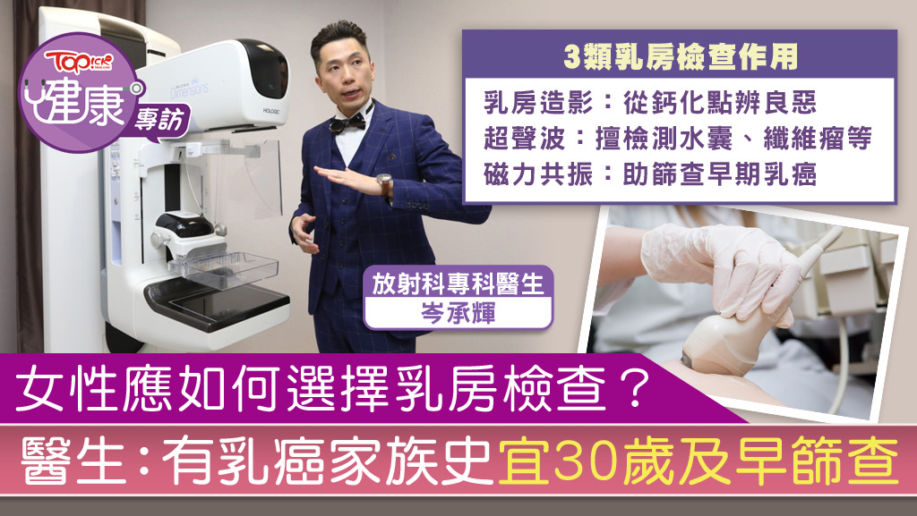
Currently, breast examinations mainly includeMammogramand ultrasound scans, which are frequently performed by specialist radiologists. Specialist radiologist Shum Shing-fai said that Hong Kong recommends that women over 40 years old undergo mammography examinations every one to two years to detect breast cancer early.
Dr. Shum pointed out that mammography should be combined with ultrasound in Oriental women to achieve better results and more comprehensive detection of cancer. Because Eastern women have denser mammary glands, the mammary glands themselves are in the form of mesh-like branches, all focused on the nipples. In 2D mammography, the breasts overlap, potentially hiding the tumor.
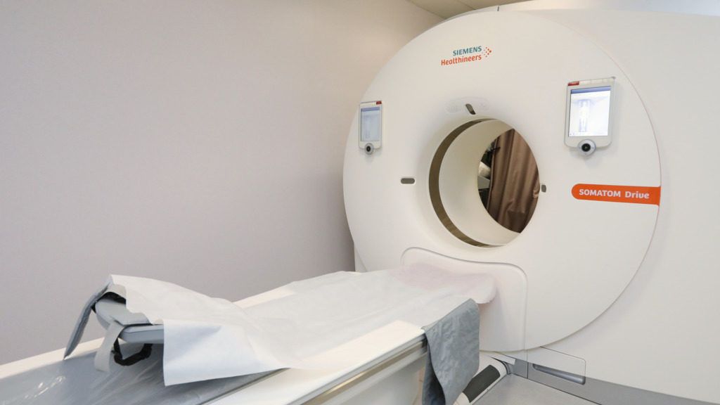
▲ 3D stereoscopic breast tomography. (Photo by Tom Ping-keung)
Newer 3D stereotactic breast tomography is seen layer by layer, as if the book is uncovered page by page, the image is clearer, higher accuracy, and can also know whether the calcification points are focused, because the focused calcification points are more likely to be malignant.
Mammography plus ultrasound
He recommended a mammogram plus ultrasound because even 3D has a chance of missing traces of the tumor. He said:
Because when some small tumors are about the same density as the mammary gland, it is possible to appear "fish that slip through the net". Coupled with ultrasound scanning, it helps to enhance detection.
Some of the tests in the market, such as thyroid ultrasound,breast examinations, lower andupper abdominal ultrasound examination, without adding X-rays, many people will ask: what is the difference between ultrasound and mammography? Dr. Sem explained that the difference is looking at different categories, both see cancerous tumors, benign tumors and fibroids, ultrasound to see water cysts or certain cancerous tumors, fibroids will be better.
Mammography is to look at some small calcification points in the breast, calcification points are divided into benign and malignant, from the mammogram can estimate whether these calcification points are malignant or benign, which are not seen by ultrasound. Therefore, having an ultrasound without a mammogram is not entirely true.
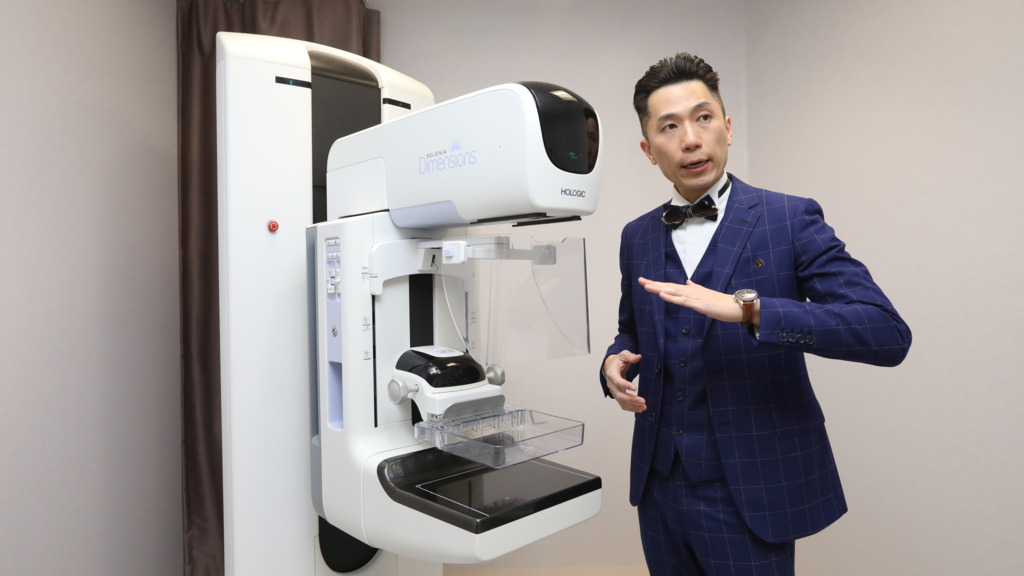
▲ Computerized CT Scan. (Photo by Tom Ping-keung)
If you have a family history, you should start checking at the age of 30 earlier
If you have a family history, you should start checking at the age of 30 earlier
A mammogram is generally not recommended at age 30 unless the patient already has a hard lump in the breast, for two reasons: First, this detection method has a certain amount of radiation. Second, young ladies have higher mammary gland density, and may not be able to find tumor traces after doing it. Early MRI is recommended unless they carry the high-risk genes BRCA-1 and BRCA-2 associated with breast cancer. Any examination has its own advantages and disadvantages, so the various methods should complement each other.
As for many people who feel that the "breast clamping" process of mammography is painful, Dr. Shum said that it has improved a lot now.
3D is not painful when using the new machine. People who have experienced it feel that it is bearable and does not even hurt, unlike the traditional 2D that makes people pinch and call for help.
Original link:https://bit.ly/3aWkiZT