
A PET scan is an advanced imaging examination that offers detailed information on a particular body organ or specific function of the body. It is commonly used to evaluate and diagnose cancer, neurological disorders of the brain, and cardiovascular disease.
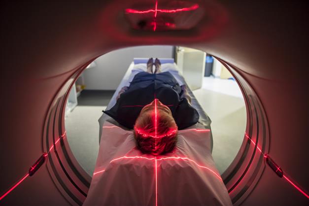
During the PET scan, a radiologist will inject a radioactive tracer into the patient’s body and images of the patient’s body are recorded with a PET scanner. A specialised camera can detect the radiation emitted by the radioactive tracer, and a multidimensional image of the examined area will be generated by the computer.
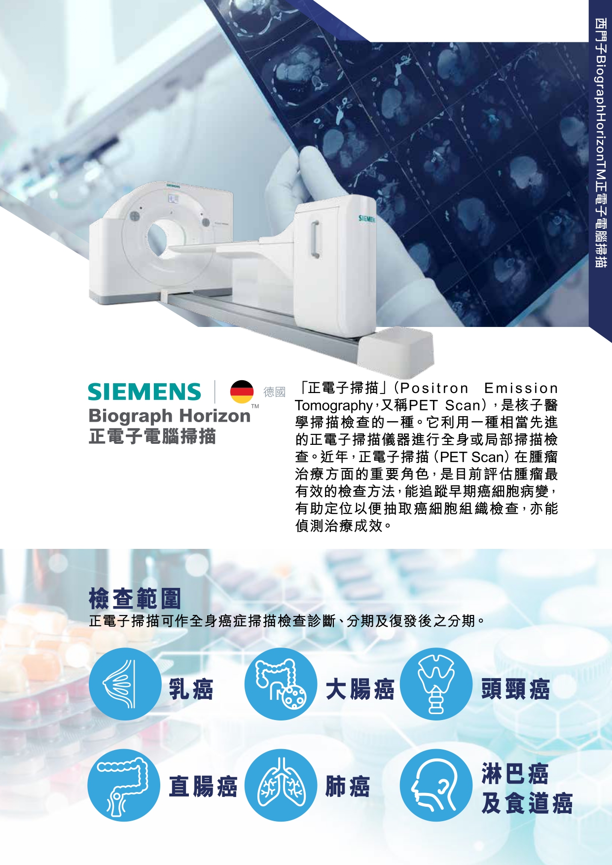
Positron Emission Tomography (PET Scan) is a form of nuclear medicine imaging. The biggest difference between a PET scan and the CT/MRI scan is their focus: the former measures metabolic activity while the latter create detailed images of the organs and tissues. Patients need to be injected with radioactive tracers (usually glucose) which will collect in tumours, inflamed areas or infections for doctors to see. If we use the analogy of photography, PET scan is like an infra-red camera while CT scan a high-definition camera. The current PET-CT scan adopted by many cancer patients combines the functional strength of PET and the structural focus of CT, hence making PET-CT a comprehensive screening. It is recommended that patients should seek medical advice from their doctor in view of the clinical signs and symptoms, so as to determine the best diagnosis.
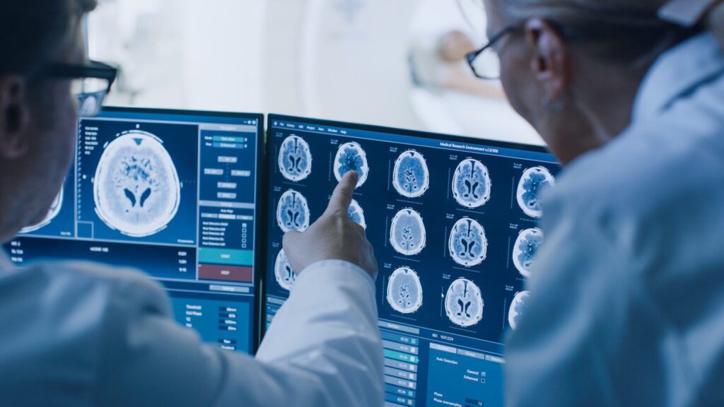
PET scans are used to detect and monitor conditions such as cancers, neurological disorders of the brain, and cardiovascular diseases. They can detect cancers before the conditions become detectable with other imaging techniques such as CT and MRI scans. PET scans also allow doctors to determine if and where cancer is spreading to other parts of the body.
PET scans can show which part of the brain is responsible for epileptic seizures, which can help doctors to develop surgery for epilepsy. It is also used to assess Alzheimer’s disease and Parkinson's disease. The images produced can pinpoint areas of the brain that are not functioning properly. Early detection of neurological disorders facilitates more effective treatment.
- Patients should not have undergone any barium x-ray examination 10 days prior to the PET-CT scan.
- A 6-hour fasting is required prior to the scan. Consumption of alcohol and beverages is prohibited (except water). Avoid medication with high sugar content (e.g. cough syrup, glucose). Avoid chewing gum or candy.
- A bland and easy-to-digest diet is recommended 1 day prior to the scan.
- Patients are advised to drink enough water, at least 6 to 8 cups of water 1 day prior to the scan.
- Avoid intense muscular activities such as strenuous exercise and heavy lifting.
- Pregnant and lactating women, people with unstable emotions, people with acutely persistent muscle cramps, and diabetic patients with poor blood sugar control should not be screened.
The radiopharmaceutical (also known as a tracer) is injected into the patient's body prior to the scan. The radiotracers usually accumulate in diseased tissues more than in healthy tissues. The images of the body are then acquired by a gamma camera in the PET scanner. The camera detects the positrons emitted from the tracers in the body, and a multi-dimensional image of the examined body part will eventually be generated by the computer. The patient has to lie still after the injection for about 1 hour to wait for the drug to be absorbed, followed by a full body scan (about 30 minutes). The patient will take another 1-1.5 hours of rest as the radiotracer becomes less radioactive over time. The risk of having side effects is low.
PET scans are rarely recommended for pregnant and breastfeeding women.
Children are more susceptible to radiation than adults and are not suitable for PET scanning.